Patient support
Our technologies
Modern diagnostics and digital fabrication make implant treatments safer, more precise and more comfortable. Here is an overview of the systems we use at the Albertine practice.
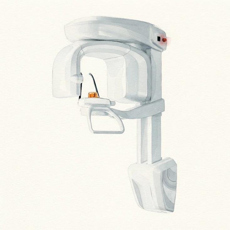
3D X-ray / CBCT
Cone-beam computed tomography produces three-dimensional jaw images with a reduced radiation dose – the basis for precise implant planning and protecting sensitive structures such as nerves and the maxillary sinus.
- CBCT significantly improves the accuracy and efficiency of implant surgery planning in the upper jaw. [1]
- Multi-source CBCT (ms-CBCT) determines bone density (Hounsfield units) more reliably than conventional CBCT. [1]
- AI-assisted CBCT analysis achieves 92–99.7% accuracy in detecting teeth and gaps. [1]
- CBCT provides higher image quality for bone structures than multidetector CT – at a significantly lower radiation dose. [2]
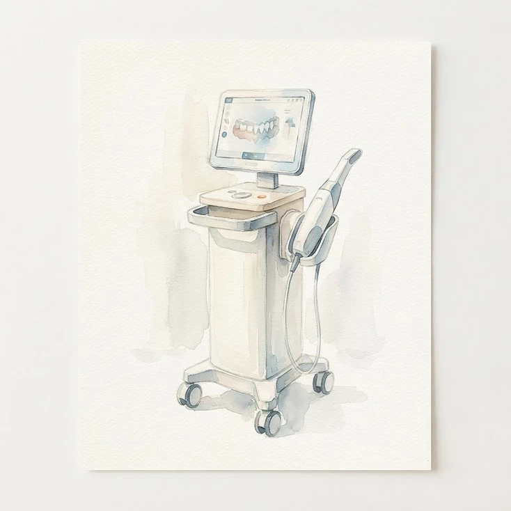
Intraoral scanners
An intraoral scanner captures your jaw digitally in seconds instead of using conventional impression material – faster, more comfortable and without gagging.
- Stereophotogrammetry-based scanners (SPG) show greater precision than conventional intraoral scanners. [3]
- The scanning strategy (quadrant versus full arch) significantly affects accuracy; lighting of at least 1,000 lux improves full-arch scans. [3]
- Digital impressions reduce treatment time and improve patient comfort. [4]
- For short-span scans (up to 4 units), digital impressions match conventional quality; full-arch scans still show variability depending on the scanner type. [4]

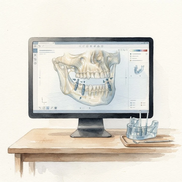
Computer-assisted implant planning
Computer-assisted planning combines CBCT data with prosthetic planning. Surgical guides produced from this data enable computer-guided, minimally invasive implant placement with high precision.
- RealGUIDE-assisted fully guided templates achieve high accuracy; longer implants show less deviation at the implant tip. [5]
- Stackable guides allow trans-sinus implant placement and sinus lifting in one step, reducing operating time in complex All-on-X cases (4 or 6 implants per jaw). [6]
- RealGuide 5.0: deviation between planned and actual implant position is less than 1 mm. [7]
- Software choice has minimal impact; the guide type (tooth-supported versus mucosa-supported) is the main factor affecting accuracy. [8]

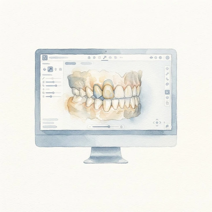
Dental CAD design
Dental CAD software converts digital scans into virtual restorations – from individual crowns to full-arch restorations. Integration with scanners, printers and milling machines makes the digital workflow seamless and efficient.
- Complete digital workflows (scan – CAD – fabrication) achieve clinically acceptable fit for fixed restorations. [9]
- Digital workflows show greater efficiency and better patient acceptance than conventional processes for implant-supported restorations. [10]
- CAD designs created with exocad achieve comparable three-dimensional accuracy and fit to conventional methods. [11]
- Digitally fabricated full dentures achieve clinically acceptable accuracy in in-vitro comparisons. [12]

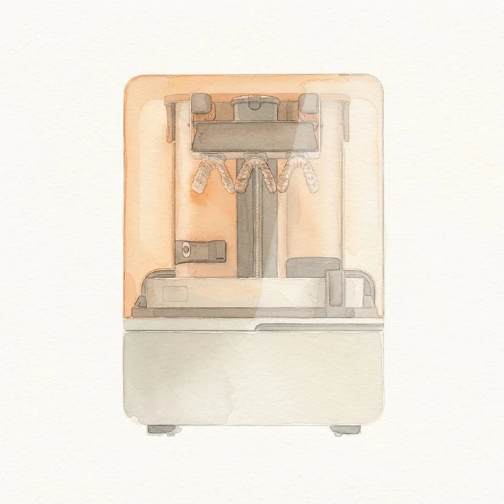
Dental 3D printing / SLA
SLA 3D printing produces highly precise working models, surgical guides, immediate restorations and permanent restorations from biocompatible resins in the dental laboratory – accurately and reproducibly.
- SLA printers show slightly higher accuracy and material efficiency, while DLP offers shorter printing times; both methods deliver clinically acceptable results. [13]
- Print orientation (0°, 10°, 20°) and shell design significantly influence dimensional accuracy and should be optimised together. [13]
- Dental-specific SLA printers are more accurate than non-dental desktop printers and are preferable for surgical guides. [14]
- Among 3D printing methods, SLA offers the best surface quality and finest detail for dental full-arch models. [15]

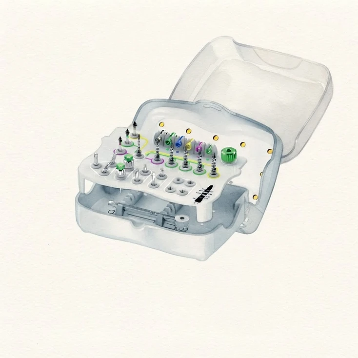
Implant systems with acid-etched surfaces
The double acid-etched surface of these implant systems is designed to support osseointegration and stable anchorage in the bone.
- DAE surfaces significantly reduce Streptococcus oralis adhesion through greater porosity, hydrophilicity and oxygen content. [16]
- Ten-year follow-up: survival above 95%, with the implant stability quotient (ISQ) remaining stable over a decade. [17]
- The DAE surface is clinically equivalent to the SLA surface (sandblasting + acid etching). [17]
- Photodynamic therapy (5-ALA gel) acts synergistically against biofilms on DAE surfaces – relevant to preventing peri-implantitis. [18]

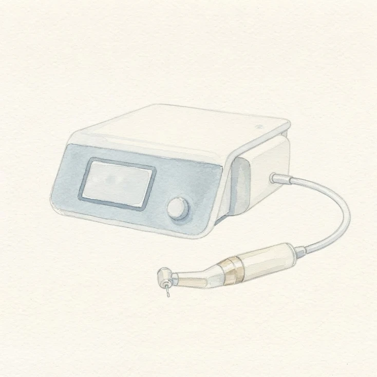
Surgical implant motors with torque analysis
Surgical implant motors with integrated torque analysis precisely control insertion speed and force – for controlled, gentle implant placement and optimal primary stability.
Implant materials
Titanium vs. zirconia – what does this mean for you?
Both materials securely anchor fixed teeth in the jaw. The choice depends on aesthetics, compatibility and your individual situation – here is an overview of the main differences.
| Feature | Titanium | Zirconia |
|---|---|---|
| Material | Metal – the established gold standard for decades[19] | High-performance ceramic (zirconia), metal-free[20] |
| Osseointegration | Very well documented, rapid bone integration[19] | Good, but with a more recent evidence base[21] |
| Survival rate | > 95% after 10 years[17] | Comparable in the short to medium term[21] |
| Biocompatibility | Excellent, bacteria-repellent surfaces[16] | Lower plaque adhesion, metal-free[20] |
| Aesthetics (front teeth) | May show a grey tint through thin gums | Tooth-coloured and natural – no grey show-through[20] |
| Metal allergy | Sensitisation is very rare | Metal-free – an alternative in cases of intolerance[20] |
| Indication | Broad range, standard treatment | Aesthetically sensitive front teeth, allergy sufferers |
| Costs | Established, economical | Usually requires greater expenditure |
Both materials are established options for implant-supported restorations. We make the final choice together in your treatment plan, taking available bone, aesthetics and compatibility into account.
Sources
Scientific sources — technologies
| # | Title | Year | Link |
|---|---|---|---|
| 1. | CBCT for Dental Implant Surgery at the Maxilla | 2026 | PMCPMC12897332 |
| 2. | CBCT Image Quality and Radiation Dose — A Systematic Review | 2025 | PMCPMC13013794 |
| 3. | Accuracy of Intraoral Scanners — A Systematic Review and Meta-Analysis | 2026 | PubMedPMID: 41093709 |
| 4. | Digital vs. Conventional Impressions — Are Intraoral Scanners Ready? (Narrative Review) | 2026 | PMCPMC12747623 |
| 5. | Influence of Implant Length on Accuracy of RealGUIDE-Guided Surgery | 2026 | PMCPMC13157714 |
| 6. | Full-Arch Trans-Sinus Implant Placement Using a Stackable Guide (RealGUIDE) | 2026 | PMCPMC13081796 |
| 7. | Prospective Clinical Study: RealGuide 5.0 .STL File Comparison | 2026 | DOIDOI: 10.3390/app16031652 |
| 8. | Computer-Guided Implant Placement Accuracy — A Systematic Review | 2026 | PMCPMC12734060 |
| 9. | The Complete Digital Workflow in Fixed Prosthodontics Updated: A Systematic Review | 2023 | PubMedPMID: 36900684 |
| 10. | Digital versus Traditional Workflows for Fabrication of Implant-Supported Rehabilitation: A Systematic Review | 2024 | PubMedPMID: 39917200 |
| 11. | Comparative Evaluation of Conventional and Digital Workflow Impressions for Implant-Supported Restorations | 2026 | PubMedPMID: 41744958 |
| 12. | Accuracy of Digital Complete Dentures: A Systematic Review of In Vitro Studies | 2021 | PubMedPMID: 32115218 |
| 13. | Comparative Evaluation of SLA and DLP 3D Printing in Dental Applications | 2025 | PMCPMC12564608 |
| 14. | Dental vs. Non-Dental SLA 3D Printers — An Accuracy Comparison | 2026 | PMCPMC11762011 |
| 15. | 3D Printing Techniques for Dental Prosthetic Models | 2026 | PMCPMC12967805 |
| 16. | The Bacterial Anti-Adhesive Activity of Double-Etched Titanium (DAE) as a Dental Implant Surface | 2020 | PMCPMC7664185 |
| 17. | Clinical Evaluation of DAE Dental Implants — A 10-Year Follow-Up | 2021 | PMCPMC8585230 |
| 18. | Photodynamic Antibiofilm Activity on DAE Titanium Surfaces (Resista) | 2022 | DOIDOI: 10.3390/biomedicines10030572 |
| 19. | Titanium Dental Implants and Osseointegration — Current Concepts and Long-term Outcomes | 2024 | PMCPMC12988001 |
| 20. | Zirconia versus Titanium Implants and the Peri-implant Soft Tissue — A Systematic Review | 2025 | PMCPMC13098211 |
| 21. | Titanium versus Zirconia Dental Implants — A Systematic Review and Meta-Analysis of Survival Rates and Marginal Bone Loss | 2026 | PMCPMC13076234 |
The cited works come from peer-reviewed specialist journals and provide scientific evidence for the content presented; they do not replace individual dental advice.